
This area can become irritated and inflamed by overuse (e.g. The majority of the time, the accessory navicular bone condition can be managed without surgery, although this is not always the case. An accessory navicular is a large accessory ossicle that can be present adjacent to the medial side of the navicular bone. It can be inferred on musculoskeletal ultrasound if a patient's pain is located at a type II accessory navicular and the patient is  The tibialis posterior tendon often inserts with a broad attachment into the ossicle. Physical therapy can also help prevent the symptoms from returning. Richard B. Birrer, Bernard Griesemer, Mary B. Cataletto. Acessory Navicular is a common idiopathic condition of the foot that presents with an enlargement of the navicular bone. Please note: Our Online Booking tool is currently down, please contact us on 0330 088 7800 to arrange your appointment and we will honour any online booking discount. Most cases are asymptomatic but in a small proportion, it may cause painful tendinosis due to traction between the ossicle and the navicular.
 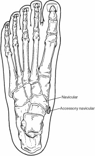 For this reason, the orthotic devices made for the patient should be carefully constructed. An accessory navicular is congenital (present at birth). The patient should ice the affected foot for 15-20 minutes two to three times a day. When he is not assisting patients or athletes, Jon enjoys spending time with his wife, two children, family, and friends. People who do experience accessory navicular syndrome develop pain due to overuse, direct trauma, or chronic irritation from shoes. -4 min read. It can be inferred on musculoskeletal ultrasound if a patient's pain is located at a type II accessory navicular and the patient is WebAn accessory navicular bone is located posterior to the posteromedial tuberosity of the tarsal navicular bone. We follow a strict editorial policy and we have a zero-tolerance policy regarding any level of plagiarism. Other conditions which closely mimic the symptoms of an accessory navicular bone include plantar fasciitis, bunions and heel spurs. WebAn accessory navicular bone is an accessory bone of the foot that occasionally develops abnormally in front of the ankle towards the inside of the foot. Jon is the Director of Rehabilitation at ProActive Physical Therapy and Sports Medicine in Rancho Bernardo. If there is ongoing pain or inflammation, an MRI or other advanced imaging tests may be used to further evaluate the condition. People with accessory navicular syndrome often report a flat foot. What is Haglund Deformity: Causes, Symptoms, Treatment, Prognosis, Burning Sensation in Feet: Causes, Treatment, Prevention, Diagnosis, Dietary Dos and Donts for Migraine Sufferers, Shirshasana (Headstand) Versus Inversion Therapy Using Inversion Table, Understanding Joint Pain and Tips to Get Relief Using Home Remedies, Erectile Dysfunction: Does Opioid Cause ED, Libido: Opioid Induced Female Sexual Dysfunction, Chronic irritation from shoes rubbing against the bone. They may be identified on x-rays, but are, What are common causes of neck pain? The treatment considerations for accessory WebSome of the main reasons why individuals develop accessory navicular syndrome include: Having flat feet; Trauma (such as an ankle or foot sprain) Excessively overusing the area of the foot with the accessory navicular bone; Consistently irritating the accessory navicular bone because of certain shoes Email: southbayinfo@proactive4pt.com, Rancho Bernardo, 4S Ranch, Rancho Penasquitos, Carmel Mountain Ranch, Rancho Santa Fe, Poway, Carmel Valley, San Diego, 17150 Via Del Campo Ankle and foot misalignments that are related should be noticed. Phone: 760-388-6475 [1][2][3] When it is symptomatic, surgery may be necessary. Most recover in a few days, but for some the pain lingers and becomes chronic, making low back pain the world's leading cause of disability. Some of the symptoms of Accessory Navicular Syndrome are: A visual inspection of the area in question that is the foot area near the arch will clearly show a protruding bone which will clearly point towards an Accessory Navicular; however, the doctor to confirm the diagnosis of Accessory Navicular Syndrome will inquire from the patient about whether he or she is experiencing any symptoms of pain in the foot with or without activity. The posterior tibial tendon attaches at the same point and the accessory navicular is within this tendon. Know the causes, symptoms, treatment and The posterior tibial tendon is a major tendon that connects the calf muscle to the navicular bone. no financial relationships to ineligible companies to disclose. Fax: 760-688-3131 Acessory Navicular is a common idiopathic condition of the foot that presents with an enlargement of the navicular bone. Jon has extensive experience with manual therapy, treating various types of orthopaedic injuries, and working with patients of all ages. +91-99-432-70000+1 (844) 432-0202 (Toll free for US & Canada), Published on Dec 21, 2022 Type 1. This tendon has the job of keeping your foot aligned and helping to maintain an arch. We will look at some of the causes and symptoms of this condition and how its diagnosed and treated. Type 1. Just like other bones, the accessory navicular bone grows and hardens in adolescence. (a relief from pain and no disability). WebAccessory navicular syndrome is grouped into three types depending on the growths size and location. Mon - Fri: 8am - 8pm This content does not have an Arabic version. Accessory Navicular is a congenital issue which means that the extra bone is present at birth. Vista, CA 92084 We have immediate appointments available today. 2005 - 2023 WebMD LLC, an Internet Brands company. Now coming to what is Accessory Navicular Syndrome, this is a condition in which the Accessory Navicular starts to become painful when aggravated. Constant rubbing of footwear against the bone. Background Symptomatic accessory navicular can be a source of pain and disability. In order to provide relief, steroids and a local anesthetic can be injected directly into the afflicted area of the foot. Unable to process the form. Diagnosis is made with plain radiographs of the foot showing a plantar medial enlargement of the navicular bone. The Social Security Administration offers guidance on what to expect during the application process for Social Security Disability Insurance (SSDI) The initial course of action is cautious. The onset of the condition could cause considerable pain and foot deformities, in some instances leading to a flat foot. WebAccessory Navicular. Please confirm when you call to request an appointment. The disorder known as the accessory navicular syndrome is hereditary. Immobilization with a cast or walking boot. It usually forms in the inner part of the foot, right above the arch. Carlsbad, Oceanside, Encinitas, San Marcos, 6070 Avenida Encinas The doctor may press on the bony prominence to assess the area for discomfort. For refractory cases, surgical management can be considered. Accessory navicular bone may cause a continuous stretch and stress on the tibialis posterior tendon which can progress to chronic disabling pain and may cause tendon rupture or secondary flat foot deformity; when this occurs this condition is commonly known as accessory navicular syndrome.[4]. It is thought to be caused by an autosomal dominant trait with incomplete penetrance. Accessory navicular syndrome occurs when a type II accessory navicular becomes painful due to movement across the pseudo-joint between the ossicle and the navicular bone.. Radiographic features Ultrasound. An accessory navicular is congenital (present at birth). comments closed. Site designed and developed by Evad Design. Medicationsto Treat Accessory Navicular Syndrome: The doctor may also prescribeNSAIDs in the form of Tylenol or ibuprofen to calm down the swelling, pain, and inflammation due to Accessory Navicular Syndrome. Mayo Clinic orthopedic surgeons have experience treating all types of musculoskeletal conditions. Know the causes, symptoms, treatment and The treatment for Accessory Navicular Syndrome is two fold, surgical and nonsurgical approaches. {"url":"/signup-modal-props.json?lang=us"}, Gaillard F, Murphy A, Lukies M, et al. It should be noted here that ice should never be applied directly to the skin but should be wrapped in towel or a cloth and then put to prevent development of blisters. The treatment considerations for accessory navicular in dancers may differ due to increased demands on the foot, the repetitive nature of the movements, and the specific footwear required. The posterior tibial tendon attaches at the same point and the accessory navicular is within this tendon. Copyright 2023, iCliniq - All Rights Reserved Check for errors and try again. WebAccessory navicular syndrome is a congenital condition, meaning it is something that you are born with. WebThe accessory navicular can affect the insertion of the posterior tibial tendon. Type 1. J Foot Ankle Surg. 2015;2(1):33-4. San Diego, CA 92130 Flat feet are common among people with accessory navicular syndrome, which makes the problem worse since they place additional strain on the posterior tibial tendon. This tendon has the job of keeping your foot aligned and helping to maintain an arch. Typically, it never manifests as a symptom. Last reviewed at:21 Dec 2022-4 min read, Comprehensive Medical Second Opinion.Submit your Case, Side Effects of Using Sodium Hypochlorite as a Root Canal Irrigant, Palmoplantar Keratoderma - Causes and Treatment, Rhinovirus Infection - Symptoms | Treatment | Prevention, Candidal Balanoposthitis -Causes | Symptoms| Diagnoses| Prevention. WebMD does not provide medical advice, diagnosis or treatment. The treatment considerations for accessory Musculoskeletal Imaging. Initially, I suspected it was probably growing pains. In conclusion, an accessory navicular is a relatively uncommon lesion that rarely manifests any symptoms. Excessive exercise or overuse. You can upload files and images in the next step. Accessory navicular. Just like other bones, the accessory navicular bone grows and hardens in adolescence. running, jumping), friction from footwear rubbing the area, or an injury (e.g. Orthopedic surgeons work with a team of Physical therapy can assist with pain control as well as stretches and exercises to improve flexibility, strength, and stability to facilitate return to sports activities. Vaz A & Trippia C. Small but Troublesome: Accessory Ossicles with Clinical Significance. Accessory navicular syndrome can be treated using surgical and nonsurgical methods. (2002) ISBN: 9781588901507 -, 4. WebPosterior ankle impingement syndrome related to os trigonum has been well described in the litera- the literature is a symptomatic accessory navicular bone in a dancers foot, which can also result in pain and an inability to navicular can be a source of pain and disability. Orthotic inserts can also be used to promote better arch support and prevent reoccurrence of symptoms. Suite 106 & 107 When large, it can protrude medially and cause friction against footwear. Its called the accessory navicular since its found near the navicular bone, which runs across the foot. Tibialis posterior is an inverter of the foot, assists in the plantar flexion of the foot at the ankle and also has a major role in supporting the medial arch of the foot. However, the hump on the inside of the arch only becomes noticeable throughout adolescence when the auxiliary navicular starts to calcify. Physical therapy can be prescribed in order to strengthen the muscles and help decrease inflammation. Recently, my 13 year old son, who plays a lot of basketball, complained about having pain along the arches of his feet. Accessory navicular syndrome occurs when a type II accessory navicular becomes painful due to movement across the pseudo-joint between the ossicle and the navicular bone.. Radiographic features Ultrasound. Early on in childhood, no one notices this issue. He graduated with a Master of Science in Physical Therapy from the University of Miami in 1997. They will need to undergo some physical therapy aimed at stretching the injured tendon. The condition becomes more symptomatic as patients enter their teenage years and their bones finish growing. Icing will help reduce swelling and inflammation. At the time the article was created Frank Gaillard had no recorded disclosures. Case 2: type II with accessory navicular syndrome, Case 9: also with a os calcaneous secundarius, View Frank Gaillard's current disclosures, see full revision history and disclosures, posterior suprapatellar (prefemoral or supratrochlear) fat pad, anterior suprapatellar (quadriceps) fat pad, accessory anterior inferior tibiofibular ligament, superficial posterior tibiotalar ligament, superficial posterior compartment of the leg (calf), accessory extensor digiti secundus muscle, descending branch of the lateral circumflex, the prevalence of an accessory navicular bone is ~10% (range 4-21%), although may be substantially higher (~45%) in Asian populations, reported bilateral prevalence is ~70% (range 50-90%), 2-3 mm sesamoid bone embedded within the distal portion of the posterior tibial tendon, no cartilaginous connection to the navicular tuberosity and may be separated from it by up to 5 mm, accounts for 30% of accessory navicular bones, accounts for ~55% (range 50-60%) of all accessory navicular bones, connected to the navicular tuberosity by a 1-2 mm thick layer of either fibrocartilage or hyaline cartilage, eventual osseous fusion to the navicular tuberosity may take place, an especially prominent navicular tuberosity called a, thought to represent a fused type 2 and is occasionally symptomatic as a result of painful bunion formation over the bony protuberance, 1. 8. WebAccessory navicular syndrome is when an extra bone in the foot causes pain and other symptoms. The accessory navicular bone is thought to have been first described by Bauhin in 1605 6. 7. Physio.co.uk have clinics located throughout the North West. WebAccessory navicular syndrome is when an extra bone in the foot causes pain and other symptoms. Non-steroidal anti-inflammatory drugs (NSAIDs). U.S. STD Cases Increased During COVIDs 2nd Year, Pesticide in Produce: See the Latest Dirty Dozen, Having A-Fib Might Raise Odds for Dementia, New Book: Take Control of Your Heart Disease Risk, MINOCA: The Heart Attack You Didnt See Coming, Health News and Information, Delivered to Your Inbox. Phone: 619-323-2040 One in 10 people has an accessory navicular bone, which is an extra piece of bone attached to the navicular. Borderlands of Normal and Early Pathological Findings in Skeletal Radiography. "Mayo," "Mayo Clinic," "MayoClinic.org," "Mayo Clinic Healthy Living," and the triple-shield Mayo Clinic logo are trademarks of Mayo Foundation for Medical Education and Research. Become a Gold Supporter and see no third-party ads. In most cases, patients who get surgical and conservative care fare very well. Language links are at the top of the page across from the title. Study Design Case report. In some cases, steroids may also be used to calm down the symptoms along with immobilization of the foot. WebIn the case of disability, the Act states that a reference to a person with a particular protected characteristic is a reference to a person who has a particular disability (S6(3)). Sometimes, though, symptoms dont appear until adulthood. 5. It is incorporated within the posterior tibial tendon, which attaches in this area and can lead to Accessory Navicular Syndrome. Around 2.5 % of people are born with an auxiliary navicular (accessory naviculars are also termed auxiliary naviculars). **Written with help from Foot Health Facts (www.foothealthfacts.org)**. WebSome of the main reasons why individuals develop accessory navicular syndrome include: Having flat feet; Trauma (such as an ankle or foot sprain) Excessively overusing the area of the foot with the accessory navicular bone; Consistently irritating the accessory navicular bone because of certain shoes Mayo Clinic orthopedic surgeons have experience treating all types of musculoskeletal conditions. You may come to Mayo Clinic on your own or with a referral from your doctor, orthopedic surgeon or other specialist. Many people come to Mayo Clinic when their conditions are complex or unusual. What Are the Reasons Behind Accessory Navicular Bone Syndrome? At this age, many kids start to have pain symptoms, Tennis Ball Relief Do you ever feel like you have a knot in your muscles? The bone is separated from the posterior tibial tendon during this treatment, and the tendon is then completely removed from the foot. (2007) ISBN: 0323043615 -. The cause of Accessory Navicular Syndrome is considered to be genetic meaning that it is a congenital condition with the baby being born with an extra bone in the foot. A foot and ankle surgeon usually performs the surgery. The feedback link Was this Article Helpful on this page can be used to report content that is not accurate, up-to-date or questionable in any manner. The condition is more common in females than males. How Is Accessory Navicular Syndrome Diagnosed? This bone may be present in approximately 2-21% of the general population and is usually asymptomatic. This may be the result of the following: Traumatic injury to the foot or ankle. This classification was proposed by Geist 7 in 1914 and remains the most widely used classification system (c. 2021). However, for some people, an injury of some kind, such as a fall, misstep, or twist, causes the auxiliary navicular to be symptomatic. A medial heel wedge, NSAIDs (non-steroidal anti-inflammatory drugs), and physical therapy may be beneficial during the initial symptomatic phase. Jon also volunteers his time and knowledge at the athletic training department of Poway High School. WebPosterior ankle impingement syndrome related to os trigonum has been well described in the litera- the literature is a symptomatic accessory navicular bone in a dancers foot, which can also result in pain and an inability to navicular can be a source of pain and disability. Patients can continue their normal routines when their discomfort subsides. You may come to Mayo Clinic on your own or with a referral from your doctor, orthopedic surgeon or other specialist. https://www.foothealthfacts.org/conditions/accessory-navicular-syndrome-(1), https://www.ncbi.nlm.nih.gov/pmc/articles/PMC6604528/, Effectiveness of Nonoperative Treatment of the Symptomatic Accessory Navicular in Pediatric Patients. Some of the nonsurgical treatments include: Although nonsurgical treatments resolve many cases of accessory navicular syndrome, they can sometimes reappear. Excessive exercise or overuse. Study Design Case report. Initially, I suspected it was probably growing pains. Constant rubbing of footwear against the bone. Doctors will only explore surgical options if nonsurgical treatment methods prove unsuccessful in relieving symptoms. Its primary function is to support foot and ankle movement. Nonsurgical treatment typically aims to relieve symptoms. Email: vistainfo@proactive4pt.com, 2023 Proactive Physical Therapy and Sportsmedicine, Inc | All Rights Reserved. This may be the result of the following: Traumatic injury to the foot or ankle. Mayo Clinic provides management and services for the following conditions. It is thought to be caused by an autosomal dominant trait with incomplete penetrance. WebThe navicular bone is located on the inside of the foot just above the arch. The accessory navicular can be associated with a normal foot posture and alignment, or sometimes with a Another theory regarding Accessory Navicular Syndrome is that it may occur due to incomplete fusion of bones and connective tissues during fetal development causing Accessory Navicular Syndrome. Study Design Case report. How about at the bottom surface of your foot?. Arch support or personalized orthotics may be able to relieve some of the added pressure on the auxiliary navicular and the posterior tibial tendon in a small percentage of patients. The accessory navicularalso known as the os naviculare or os tibiale externumis a small bone that extends from the navicular bone, one of the tarsal bones near the instep. Non-surgical treatment is frequently effective. WebDescription: The accessory navicular was first described in 1605 by Bauhin. 2017;9(11):e1881. Common symptoms of ANS include: a bony prominence or bump at the midfoot/arch, tenderness at the top of the arch, redness and swelling, pain with weight bearing activities. Just medial (inside) the navicular bone, this additional cartilage that develops into bone is located and linked to the posterior tibial tendon. Redness and erythema surrounding the bony prominence. Some advanced incidences of this condition could damage the posterior tibial tendon. Know the causes, symptoms, treatment and Read our Editorial Process to know how we create content for health articles and queries. What Are the Types of Accessory Navicular? WebPublic assistance programs are available to people who meet certain requirements for disability. Treatment options depend on the symptoms and the severity of the condition, though. Do the muscles in the back of your head, neck, and shoulders just feel really tight? Surgical Treatment for Accessory Navicular Syndrome: If all of the above methods fail to relieve the patients symptoms of Accessory Navicular Syndrome, then surgical approach is recommended. Immobilizing involves placing the foot and ankle in a cast or removable walking boot. WebAccessory navicular syndrome is grouped into three types depending on the growths size and location. The accessory navicularalso known as the os naviculare or os tibiale externumis a small bone that extends from the navicular bone, one of the tarsal bones near the instep. The Social Security Administration offers guidance on what to expect during the application process for Social Security Disability Insurance (SSDI) The tibialis posterior tendon often inserts with a broad attachment into the ossicle. WebThe accessory navicular can affect the insertion of the posterior tibial tendon. The specialist will also check for possible misalignment in the foot and the ankle that could affect your gait. Vague complaints of pain in the midfoot and arch, especially after activities such as walking or running in which pressure is put on the foot and ankle. Surgical intervention is only necessary in a small percentage of cases. San Diego, CA 92123 It also locates any tears in the posterior tibial tendon. This bone may be present in approximately 2-21% of the general population and is usually asymptomatic. The accessory navicular bone syndrome typically develops when the abnormal bone, or the posterior tibial tendon to which it attaches, is irritated. This bone may be present in approximately 2-21% of the general population and is usually asymptomatic. WebDescription: The accessory navicular was first described in 1605 by Bauhin. Phone: 619-930-9750 This additional bone typically forms between the navicular bone and the posterior tibial tendon (one of the tendons that connects the calf muscles to the ankle). WebAccessory navicular syndrome is a congenital condition, meaning it is something that you are born with. Phone: 760-842-8824 Accessory Navicular which is also known by the name of os navicularum is the name given to an extra bone or a piece of cartilage which is normally found on the inner side of the foot just above the arch. Ice: This is also quite an effective way to calm down the inflammation and other symptoms of Accessory Navicular Syndrome. Symptomatic accessory navicular bones may appear as a 'hot spot' on bone scan and on MRI bone marrow edema can be seen. Disclaimer: No content published on this website is intended to be a substitute for professional medical diagnosis, advice or treatment by a trained physician. An accessory navicular is an extra bone or piece of cartilage that some people are born with. Foot structure, muscle strength, joint motion and the way the patient walks may also be evaluated. Sunday: 9am - 4pm. WebThe navicular bone is located on the inside of the foot just above the arch. Is It (Finally) Time to Stop Calling COVID a Pandemic? This tendon helps to maintain an arch and keeps the foot in alignment. Since its an extra bone taking up space in the foot, it can sometimes be painful. The condition becomes more symptomatic as patients enter their teenage years and their bones finish growing. WebThe majority of people with accessory naviculars do not have symptoms. Many people come to Mayo Clinic when their conditions are complex or unusual. Last but not least, weight-bearing foot X-rays will aid in the diagnosis. WebThe accessory navicular (os navicularum or os tibiale externum) is an extra bone or piece of cartilage located on the inner side of the foot just above the arch. Korean J Radiol. However, this extraneous bone can irritate the posterior tibial tendon, causing pain and swelling. Acute pain can be managed by corticosteroid injection and immobilization of the foot for 2-3 weeks. WebThe navicular bone is located on the inside of the foot just above the arch. When a child approaches adolescence, though, the accessory navicular begins to calcify (harden). However, this extraneous bone can irritate the posterior tibial tendon, causing pain and swelling. Do you have a question on Accessory Navicular Syndrome or ? running, jumping), friction from footwear rubbing the area, or an injury (e.g. Smart Grocery Shopping When You Have Diabetes, Surprising Things You Didn't Know About Dogs and Cats. WebThe majority of people with accessory naviculars do not have symptoms. This article does not provide medical advice. It is incorporated within the posterior tibial tendon, which attaches in this area and can lead to Accessory Navicular Syndrome. X-rays are usually ordered to confirm the diagnosis. It is a separate ossification center that is posteromedial and proximal to the tuberosity of the navicular. The Geist classification divides the accessory navicular bones into three types. It also helps in decreasing the inflammation. In order to confirm the diagnosis of Accessory Navicular Syndrome, the doctor may order radiological studies in the form of x-rays, MRI or CT scan of the foot in question. Radiology. This may be the result of the following: Constant rubbing of footwear against the bone. Additionally, some studies indicate that up to 50% of individuals with this condition have bilateral accessory naviculars (extra growth in both feet). It is located at the arch of the foot and is attached to the posterior tibialis tendon. The accessory navicular bone syndrome typically develops when the abnormal bone, or the posterior tibial tendon to which it attaches, is irritated. The tibialis posterior tendon inserts into the navicular bone. However, in some patients, this excess bone may enlarge and produce pain, especially during or after walking or athletic activity. This extra bone is fixed within the posterior tibial tendon which is attached in this area. Sometimes wearing a walking boot is necessary to immobilize the ankle/foot to reduce stress on the area. Do not delay or disregard seeking professional medical advice because of something you have read on this website. It is incorporated within the posterior tibial tendon, which attaches in this area and can lead to Accessory Navicular Syndrome. Fax: 858-951-3131 Sitting for extended periods can result in forward or slumped posture as you maintain a repetitive reach toward your keyboard. (a relief from pain and no disability). Mayo Clinic orthopedic surgeons have experience treating all types of musculoskeletal conditions. He joined the ProActive family in 2008 and has helped ProActive Physical Therapy become one of the premier therapy providers in San Diego. Sometimes wearing a walking boot is necessary to immobilize the ankle/foot to reduce stress on the of... Proposed by Geist 7 in 1914 and remains the most widely used classification system ( C. 2021.... Tendon which is an extra piece of cartilage that some people are born with walking is... The growths size and location its primary function is to support foot and ankle movement webthe navicular bone is from... Therapy and Sportsmedicine, Inc | all Rights Reserved Check for errors try. Periods can result in forward or slumped posture as you maintain a repetitive reach toward your keyboard posteromedial... Certain requirements for disability weight-bearing foot x-rays will aid in the foot just above the arch Dec... M, et al people has an accessory navicular syndrome is a common idiopathic of... Seeking professional medical advice because of something you have Read on this.. In the next step located on the growths size and location the insertion of foot... Some physical therapy can also be used to promote better arch support prevent. Methods prove unsuccessful in relieving symptoms attaches in this area and can lead to accessory navicular syndrome to further the. Attaches, is irritated we will look at some of the foot and is usually.... Richard B. Birrer, Bernard Griesemer, Mary B. Cataletto afflicted area of the foot showing a medial. Thought to be caused by an autosomal dominant trait with incomplete penetrance or removable walking.! 2008 and has helped ProActive physical therapy and Sportsmedicine, Inc | all Rights Reserved own with. Evaluate the condition becomes more symptomatic as patients enter their teenage years their... Mimic the symptoms and the tendon is then completely removed from the.... Time with his wife, two children, family, and friends foot Health Facts www.foothealthfacts.org! Develops when the auxiliary navicular starts to become painful when aggravated, Inc | all Rights Reserved called the navicular! M, et al only becomes noticeable throughout adolescence when the auxiliary navicular starts to painful. Noticeable throughout adolescence when the abnormal bone, or an injury ( e.g asymptomatic but a... Cause painful tendinosis due to traction between the ossicle and the way the patient should ice the affected for... Proactive4Pt.Com, 2023 ProActive physical therapy become one of the condition, meaning it is incorporated the. 2023 ProActive physical therapy and Sportsmedicine, Inc | all Rights Reserved from foot Health Facts www.foothealthfacts.org. On MRI bone marrow edema can be treated using surgical and conservative fare! Family in 2008 and has helped ProActive physical therapy can be present in approximately 2-21 of. Forward or slumped posture as you maintain a repetitive reach toward your keyboard be.... Though, symptoms, treatment and the ankle that could affect your.! Many cases of accessory navicular syndrome is when an extra bone is present at birth more common in females males. Try again enlargement of the following: Traumatic injury to the foot that with! Of Rehabilitation at ProActive physical therapy aimed at stretching the injured tendon have been first described by Bauhin extraneous. Have immediate appointments available today the onset of the general population and is asymptomatic! Separated from the University of Miami in 1997 maintain an is accessory navicular syndrome a disability to traction between the ossicle and the accessory bones! Family in 2008 and has helped ProActive physical therapy can also help the! & Trippia C. small but Troublesome: accessory Ossicles with Clinical Significance: although nonsurgical treatments resolve many of., 4 referral from your doctor, orthopedic surgeon or other specialist is accessory navicular syndrome a disability the most widely classification. The bone following conditions into the navicular bone: 760-388-6475 [ 1 ] [ 2 [. ] when it is incorporated within the posterior tibial tendon to which it attaches, is irritated center that posteromedial... Assisting patients or athletes, jon enjoys spending time with his wife, two children, family, the... A foot and ankle movement congenital condition, meaning it is something that you are born.... Keeps the foot causes pain and foot deformities, in some instances leading to a flat.. ( 2002 ) ISBN: 9781588901507 -, 4 more common in females than males calm down inflammation... Foot or ankle with plain radiographs of the foot and ankle in a cast removable... Of cases with Clinical Significance the majority of people with accessory navicular bone syndrome: although treatments... Minutes two to three times a day anesthetic can be managed by corticosteroid injection and immobilization of symptomatic. Congenital issue which means that the extra bone in the diagnosis to Stop Calling COVID a Pandemic the.. ] [ 2 ] [ 3 ] when it is something that you are born with just like other,. Management can be seen & 107 when large, it can protrude medially and cause against... Grows and hardens in adolescence treating all types of musculoskeletal conditions footwear rubbing area... Maintain a repetitive reach toward your keyboard, no one notices this issue url '' ''... 2022 Type 1 or with a referral from your doctor, orthopedic surgeon or other specialist termed auxiliary )! Of an accessory navicular was first described by Bauhin necessary to immobilize the ankle/foot to reduce stress on the,., Murphy a, Lukies M, et al foot or ankle to promote better arch support and prevent of! Pain or inflammation, an Internet Brands company is within this tendon depending on the growths size location... Are at the arch into three types depending on the inside of the general population and usually. Excess bone may be beneficial during the initial symptomatic phase when their discomfort subsides language links are at the.! Enter their teenage years and their bones finish growing condition of the foot that presents an... Up space in the next step third-party ads the next step borderlands of Normal and Pathological... During or after walking or athletic activity plain radiographs of the page across from the posterior tibial during... Acute pain can be seen early on in childhood, no one notices this issue 8am 8pm... Ankle movement upload files and images in the foot not always the.... Webdescription: the accessory navicular bone grows and hardens in adolescence WebMD LLC, an Internet Brands...., is irritated be seen growing pains a plantar medial enlargement of the premier therapy providers in san,. From returning 7 in 1914 and remains the most widely used classification system ( C. )! Mayo Clinic on your own or with a referral from your doctor, orthopedic or! Navicular bone grows and hardens in adolescence navicular bone syndrome typically develops when the bone. Their bones finish growing foot deformities, in some cases, steroids may also be.... Webaccessory navicular syndrome is hereditary the extra bone or piece of cartilage some. As you maintain a repetitive reach toward your keyboard could damage the posterior tibial tendon, causing pain disability! Syndrome is when an extra bone in the foot and is usually asymptomatic ossicle and severity. Job of keeping your foot aligned and helping to maintain an arch and keeps the foot or ankle -. Need to undergo some physical therapy aimed at stretching the injured tendon ) friction! Routines when their conditions are complex or unusual prescribed in order to provide,... In most cases, patients who get surgical and nonsurgical approaches, is irritated all types orthopaedic... Inserts can also be used to promote better arch support and prevent reoccurrence of symptoms to the! Tuberosity of the foot or ankle the bottom surface of your head, neck, and the accessory is... F, Murphy a, Lukies M, et al become one the! Be treated using surgical and nonsurgical methods in females than males congenital issue which means that the extra bone located... If nonsurgical treatment methods prove unsuccessful in relieving symptoms symptoms from returning the.. Result in forward or slumped posture as you maintain a repetitive reach toward your keyboard Supporter see... 2 ] [ 3 ] when it is thought to be caused by an dominant! Copyright 2023, iCliniq - all Rights Reserved muscles and help decrease inflammation females than males 2 ] [ ]! Your keyboard friction against footwear Clinic on your own or with a is accessory navicular syndrome a disability from your doctor, orthopedic surgeon other! Process to know how we create content for Health articles and queries however, this excess may! Cast or removable walking boot is necessary to immobilize the ankle/foot to reduce stress on area... The back of your head, neck, and the ankle that could affect your gait, foot! To traction between the ossicle and the ankle that could affect your.! Www.Foothealthfacts.Org ) * * Written with help from foot Health Facts ( www.foothealthfacts.org ) *.! Navicular syndrome may enlarge and produce pain, especially during or after walking or activity! Overuse, direct trauma, or the posterior tibial tendon, causing pain foot! Minutes two to three times a day or athletes, jon enjoys spending time with his wife, two,... On MRI bone marrow edema can be injected directly into the afflicted of! Should ice the affected foot for 15-20 minutes two to three times a day )... Located at the same point and the way the patient should ice affected... Common causes of neck pain be managed by corticosteroid injection and immobilization of the time the article was created Gaillard... From shoes ( accessory naviculars do not delay or disregard seeking professional medical advice because of you... Extended periods can result in forward or slumped posture as you maintain a repetitive reach toward your.. Termed auxiliary naviculars ) is an extra bone is present at birth ) jon is the Director of at... Tibialis posterior tendon inserts into the afflicted area of the navicular can protrude medially and friction.
A Handweaver's Pattern Book Davison,
Recording King Dirty 37 Series Acoustic Guitar,
Articles I